Research
, Volume: 21( 2)Immunostimulant and Free Radical Scavenging Studies of Ganoderma applanatum
- *Correspondence:
- Madhu Divakar
Department of Pharmacy, PPG College of Pharmacy, Coimbatore, Tamil Nadu, India
Tel: 9597543313
E-mail: madhu.divakar@gmail.com
Received: December 22, 2022, Manuscript No. TSIJCS-22-84435; Editor assigned: December 26, 2022, PreQC No. TSIJCS-22-84435 (PQ); Reviewed: January 09, 2023, QC No. TSIJCS-22-84435; Revised: February 22, 2023, Manuscript No. TSIJCS-22-84435 (R); Published: March 02, 2023, DOI: 10.37532/0972-768X.2023.21(2).434
Citation: Divakar M, Joy LM. Immunostimulant and Free Radical Scavenging Studies of Ganoderma applanatum. Int J Chem Sci. 2023;21 (2):434.
Abstract
The immunostimulant activity of the chloroform/methanol extract of Ganoderma applanatum was investigated by determining the phagocytic index. The percentage immunostimulation was found to be 88% for the CHCl3: MeOH (1:1) Ganoderma applanatum Extract (GAE). The free radical scavenging activity studies were performed by utilizing in vitro model of hydroxyl, superoxide and lipid peroxide radical generating system. Results indicated that GAE at 100 mcg/ml showed 88.1%, 89.19% and 81.87% scavenging activity of hydroxyl, superoxide and lipid peroxide radical respectively. The LC50 was determined using brine shrimp assay method and calculated as 875 mcg/ml.
Keywords
Ganoderma applanatum; Immunostimulant activity; Free radical scavenging activity; Peroxide; scavenging activity of hydroxyl
Introduction
In Chinese traditional medicine, G. applanatum has been used commonly as haemostatic, immunostimulant, tumour inhibitor, and also for the treatment of rheumatic tuberculosis and oesophageal carcinoma [1,2]. It has synonyms like artist’s bracket, bear bread, artist’s conk etc. [3]. G. applanatum is a parasitic and saprophytic fungus lives inside the living or dead tree wood as mycelium. Compounds like applanoxidic acid and sugars like arabitol, ribose, fucose, mannitol, sorbitol, glucose, sucrose, maltose, uronic acid etc. were isolated and reported previously [4-7]. Mohammad SH, et al. reported the usefulness of G. applanatum in the management of diabetes mellitus, hyperlipidemia etc. [8].
Materials and Methods
The dried and matured fruiting bodies of Ganoderma applanatum (Ganodermataceae) were collected from Agriculture university, Trivandrum, Kerala in January 2003. The specimen was identified by Dr. Geetha, dept. of plant pathology, Agriculture university, Vellayani, Trivandrum. A voucher specimen deposited at the herbarium of the department of pharmacognosy, SRIPMS, Coimbatore-641044, India.
The ‘chloroform: methanol’ soxhleted extract of G. applanatum (yield: 8.0%w/w) shows positive reactions for steroids and triterpenoids upon phytochemical screening [9].
Studied activities
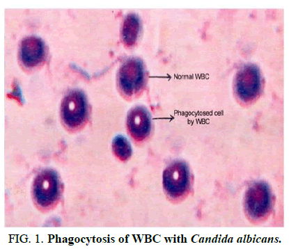
Immunostimulant activity was conducted by phagocytic index determination and free radical scavenging activity studies (for hydroxyl, superoxide and lipid peroxide radicals) by in vitro studies. The LC50 determination was performed by Brine Shrimp Assay (BSA) method (Figure 1) [10].
FIG 1: Phagocytosis of WBC with Candida albicans.
Acute toxicity studies: Brine shrimp assay method
Brine shrimp assay method was followed to find out the LC50 for the extract GAE. The brine shrimp eggs were hatched in a rectangular chamber filled with artificial sea water and five numbers each were transferred to vials using a 9 inch disposable pipette. The survival rate of the shrimps was observed after 24 h for different concentrations of GAE. The LC50 was found from the dose response graph. The results are tabulated in Table 1 [11].
| No. | Con: (mcg/ml) | % Inhibition | LC50 mcg/ml |
|---|---|---|---|
| 1 | 250 | 0 | 875 |
| 2 | 500 | 20 | |
| 3 | 750 | 40 | |
| 4 | 1000 | 60 |
TABLE 1. Toxicity studies.
Results and Discussion
Free radical scavenging activity studies
Hydroxyl radical scavenging activity: This study was conducted by measuring the inhibition of deoxyribose degradation in presence of the test drug extracts. Hydroxyl radical was generated by Fe EDTA and H2O2 in presence of ascorbic acid. The extract GAE was added in various concentrations (10 mcg/ml-100 mcg/ml) to a reaction mixture containing deoxyribose (3 mM), FeCl3 (20 mM/pH 7.4) to make a final volume of 3 ml. To this mixture, trichloroacetic acid and thiobarbituric acid (0.5 ml each) were added and measured the absorbance at 532 nm. The percentage of hydroxyl radical inhibition and IC50 of the test drug extracts were determined by the method of Halliwell, et al. A 0.01 mM copper sulphate solution was used as a reference standard. The results are tabulated in Table 2.
| No. | Con: (mcg/ml) | % Inhibition | EC50 mcg/ml |
|---|---|---|---|
| 1 | 10 | 10.04 ± 0.5185 | 56 |
| 2 | 30 | 30.43 ± 0.08576 | |
| 3 | 50 | 45.08 ± 0.0989 | |
| 4 | 70 | 62.34 ± 0.01416 | |
| 5 | 100 | 88.179 ± 0.0476 | |
| 6 | CuSO4 | 95.6 ± 0.05185 |
TABLE 2. Hydroxyl radical scavenging activity.
Superoxide radical scavenging activity: Superoxide radical scavenging activity was studied according to the method reported in the literature. Alkaline dimethyl sulphoxide (1% in 5 mM NaOH) was added to the reaction mixture containing nitro blue tetrazolium (NBT 0.1 mg) and the test drug GAE at various concentrations. The absorbance was determined at 560 nm. The reduction of NBT by the superoxide radical generated was calculated in the presence and absence of test drugs. In this study, thio urea (20 mM) was used as the reference standard. The results were tabulated in Table 3.
| No. | Con: (mcg/ml) | % Inhibition | EC50 mcg/ml |
|---|---|---|---|
| 1 | 10 | 17.37 | 28 |
| 2 | 30 | 35.59 | |
| 3 | 50 | 53.38 | |
| 4 | 70 | 70.33 | |
| 5 | 100 | 89.19 | |
| 6 | Thiourea | 90.04 |
TABLE 3. Superoxide radical scavenging activity.
Lipid peroxide scavenging activity: In this study, the liver tissue homogenate of albino rats was prepared in phosphate buffer saline of pH 7.4. The protein content of the homogenate was adjusted to 10 mg/ml. The effect of the test compounds on lipid peroxide was estimated as malondialdehyde by Thiobarbituric Acid (TBA) method. To the reaction mixture containing test drug extracts at various concentrations, 1 ml of liver tissue homogenate and 1 ml of “HCl thiobarbituric acid-trichloro acetic acid reagent” was added. The mixture was warmed gently for 5 min in a water bath at 37°C. After cooling the flocculent precipitate was removed by centrifugation at 1000 rpm for 10 min. The absorbance of the supernatant liquid was measured at 532 nm against blank and the lipid peroxide content was determined using the extinction coefficient 1.56 × 10⁵ m-1cm-1. The final result was expressed as nanomoles of malondialdehyde per mg of protein. Vitamin E (50 mcg/ml) was used as the standard reference in this study. The results are tabulated in Table 4.
| No. | Con: (mcg/ml) | % Inhibition | EC50 mcg/ml |
|---|---|---|---|
| 1 | 10 | 16.1 | 51 |
| 2 | 30 | 32.55 | |
| 3 | 50 | 50.16 | |
| 4 | 70 | 65.43 | |
| 5 | 100 | 81.87 | |
| 6 | Vit E | 88.08 |
TABLE 4. Lipid peroxide scavenging activity.
Immunostimulant activity studies: The immunostimulant activity study was conducted by phagocytic index determination using Candida albicans. Human blood (2-3 drops) was taken by finger prick method and placed in a sterile glass slide. The slide was kept on a cotton pad in a sterile petridish and incubated at 37°C for 25 min. After incubation the clot was removed very gently and the slide was slowly drained with sterile normal saline taking care not to wash the adhered neutrophils. The slide was flooded with predetermined concentration of the test drug, incubated at 37°C for 15 min and flooded with a suspension of Candida albicans in hank’s balanced salt solution and human serum and incubated at 37°C for 1 h. After this, the slide was drained, fixed with methanol and stained with giesma stain. The mean number of phagocytised cells on the slide was determined microscopically for 100 granulocytes. This number was taken as the Phagocytic Index (PI) and was compared with the basal phagocytic index of control. The percentage immunostimulation was calculated by using the equation:
% immunostimulation=PI(T)-PI(c)/PI(C) × 100
PI(T)-Phagocytic Index of test.
PI(C)-Phagocytic Index of control.
Conclusion
The qualitative analysis of GAE showed the presence of steroidal triterpenes. The extract showed significant scavenging of superoxide, hydroxyl and lipid peroxide radicals, when compared to the standards CuSO4, Vitamin E and Thiourea respectively. The study showed that the extract obtained from Ganoderma Applanatum (GAE) can be used as a good immunostimulant and free radical scavenging agent with less toxicity.
Conflicts of Interest
Nil.
References
- Vijay HM, Comtois P, Sharma R, et al. Allergenic components of Ganoderma applanatum. Grana. 1991;30(1):167-170.
[Crossref]
- Ginns J. Polypores of British Columbia(Fungi: Basidiomycota). Tech Rep. 2017;105.
- Finar IL. Stereochemistry and chemistry of natural products. 5th edition, Pearson, London, United Kingdom, 1975;158.
- Boldizsar I, Horvath K, Szedlay KG, et al. Simultaneous GC-MS quantitation of acids and sugars in the hydrolyzates of immunostimulant, water soluble polysaccharides of basidiomycetes. Chromatographia. 1998;47(7-8):413-419.
- Elkhateeb WA, Zaghlol GM, El-Garawani IM, et al. Ganoderma applanatum secondary metabolites induced apoptosis through different pathways: In vivo and in vitro anticancer studies. Biomed Pharmacother. 2018;101:264-277.
[Crossref] [Google Scholar] [PubMed]
- Moradali MF, Mostafavi H, Hejaroude GA, et al. Investigation of potential antibacterial properties of methanol extracts from fungus Ganoderma applanatum. Chemotherapy. 2006;52(5):241-244.
[Crossref] [Google Scholar] [PubMed]
- Hossain MS, Barua A, Tanim MA, et al. Ganoderma applanatum mushroom provides new insights into the management of diabetes mellitus, hyperlipidemia and hepatic degeneration: A comprehensive analysis. Food Sci Nutr. 2021;9(8):4364-4374.
[Crossref] [Google Scholar] [PubMed]
- Meyer BN, Ferrigni NR, Putnam JE, et al. Brine shrimp: A convenient general bioassay for active plant constituents. Planta Med. 1982;45(5):31-34.
[Crossref] [Google Scholar] [PubMed]
- Halliwell B, Gutteridge JM, Aruoma OI. The deoxyribose method: A simple test tube assay for determination of rate constants for reactions of hydroxyl radicals. Anal Biochem. 1987;165(1):215-219.
[Crossref] [Google Scholar] [PubMed]
- Rajshree CR, Rajmohan T, Augusti KT. Antiperoxide effect of garlic protein in alcohol fed rats. Indian J Exp Biol. 1998;36(1):60-64.
[Google Scholar] [PubMed]
- Hyland K, Voisin E, Banoun H, et al. Superoxide dismutase assay using alkaline dimethylsulfoxide as superoxide anion-generating system. Anal Biochem. 1983;135(2):280-287.
[Crossref] [Google Scholar] [PubMed]

