Research
, Volume: 14( 5)Evaluation of Protective Effects of Moringa Leaves Extract on BM Cells Injury Caused by Radiation Using Thermodynamic State Functions Derived Dielectric Parameters
- *Correspondence:
- Mohamed MM Elnasharty , Microwave Physics Department, Physics Division, National Research Centre, -33 El Bohouth St., Dokki, Giza, Egypt, E-Mail: mohamed.elnasharty@gmail.com
Received: October 09, 2018; Accepted: October 26, 2018; Published: October 30, 2018
Citation: Elnasharty MMM and Elwan AM. Evaluation of Protective Effects of Moringa Leaves Extract on BM Cells Injury Caused By Radiation Using Thermodynamic State Functions Derived Dielectric Parameters. Biotechnol Ind J. 2018;14(5):175.
Abstract
Using a natural supplement Moringa oleifera leaves extract (MOLE) to enforce the bio system struggling against irradiation damage is the issue addressed in this work. We performed the experiment on Wistar albino rats and had our measurements on the bone marrow extracted from the femur bone. New physical parameters are presented to investigate the effects of both irradiation and the counter effect of the bio system enforced by the Moringa leaves extract. Most of the physical parameters used in the study is dielectric and its related thermodynamic state functions expressed an opposite behavior to that reported by irradiation during the period of the extract administration.
Keywords
Moringa leaves extract; Bone marrow; Radiation; Dielectric parameters; Thermodynamic state functions
Introduction
Phosphorus (P) is an essential element required for plant growth and is involved in many plant metabolic functions [1,2]. Production of blood cells depends on the hematopoietic stem cells of bone marrow. Hematopoietic stem cells produce all types of blood cells such as red and white blood cells, platelets, granulocytes, B and T-lymphocytes and macrophages [1].
Bone marrow failure leads to an inability of producing the blood cells. Several different reasons can cause bone marrow failure syndromes such as exposure to radiation, chemicals, viruses, or toxins, which lead to anemia and depletion in the immune system [2]. The stem cells of bone marrow can be destroyed by gamma radiation (0.5-2Gy) as they possess a high sensitivity against ionizing radiation [3].
Numerous synthetic and natural materials, such as melatonin, substance P, flavonoids and Mentha piperita have been tested to provide a protection of BM against the radiation damage [4-7]. Also, Moringa oleifera plant is one of the best plants that help in the protection against radiation damage [8-11]. In this study, this plant has been chosen to protect bone marrow cells against radiation and study this effect on the thermodynamic properties of bone marrow.
Moringa oleifera is considered a medicinal plant used in many countries such as India and China. Leaves of this plant contain a vast amount of nutrients like essential and non-essential amino acids, vitamins and minerals. The dried leaves contain higher concentrations of nutrients than fresh leaves, because of water loss, except for vitamin C as it is affected by leaves dryness [12,13]. Additionally, the phytochemical analysis of Moringa leaves shows that they contain tannins, flavonoids, Saponins, alkaloids and glycosides, therefore, Moringa leaves have anti-bacterial, anti-parasitic, anti-viral and antiinflammatory activities [14-16].
Bone marrow is the most sensitive tissue within the body to radiation. The less differentiated the cells the more sensitive they are to radiation. The problem of accidentally irradiated subjects is to determine or estimate the dose they were exposed to as early as possible to be able to enforce the subject with suitable molecules to be used for both fightings and repairing the induced damage. Waiting till the symptoms to appear would, in serious cases, be too late. Consequently, biochemical markers and pathological examinations which express significant values only after the death or damage of concerned cells make it too late to interfere if we are looking for damage prevention. That was the reason for us to think of superior biophysical markers that could present better and faster estimators for different radiation doses and dose rates. Using both dielectric parameters and thermodynamic state functions derived from them as biophysical markers. Dielectric parameters are very sensitive to any electrical inhomogeneity, changing the net dipole moment, and their dependent thermodynamic state functions of the measured sample as proven in our previous work [11,17-19]. This work continues the investigation of the effect of Moringa leaves extract on the bone marrow of total body irradiated female Wistar albino rats through their thermodynamical state functions derived from dielectric data of bone marrow.
In this work, we used 168 female rats as we did not want the study to be subjected to other parameters that will not be taken into consideration such as different response due to the difference in animal type. Moreover, the dose of moringa extract used during the course of our work (1g/kg) was determined upon reading the effects and response of animals to wide dose range used by other researchers, ranged from 300 mg/kg to 5.0 gm/kg of animal’s body weight. Such a wide range of MOLE was reported to be safe in these references [8-17,19]. However we, authors, noticed from others’ work that there was a stress effect for MOLE on the animals which appeared in the results. Upon these facts, we decided to use a daily medium dose of 1.0 gm/kg body weight.
The subsequent equations use dielectric measurements of bone marrow from our previous paper [11] where rats were subjected to a total dose of 7 Gy at a dose rate of 325.89 mGy/min. The first equation estimates the activation energy of the detected process during impedance measurements of bone marrow in our previous work [11]. The value of activation energy, Ea, can be deduced from relaxation time, equation (1), or by replacing the relaxation time, τ, by the direct current conductivity, the value of the conductivity which is frequency independent.
 (1)
(1)
Where T is the absolute temperature, R is gas constant and  is a pre-exponential factor [20].
Activation energy means the least energy amount required to do the process of interest. This definition would assist the researcher to track the studied process during different stages of experiment consequently permitting understanding the system status.
The enthalpy change, ΔH, a state function and is derived from the relaxation time of the assessed process.
 (2)
(2)
Where, ΔH is the enthalpy change estimates heat and/or work transfer between the system studied and its surrounding [21]. Estimation of ΔH would reveal the net result of the relation between the system and its surrounding under the effect of the used effector, irradiation in this case which delivers information about the complications occurred within the system from the effector.
Helmholtz free energy is a state function expresses the amount of energy that is free for the system to use. Assessing the change in this type of energy forms an impression of the system under study and the way it is affected by parameters studied.
 (3)
(3)
Where,  , are the Helmholtz free energies as functions of absolute temperature, T, when applying the electric
field, E, or switching it off respectively, εo is the permittivity of free space and εs is the static permittivity, the part of the
permittivity spectrum where the permittivity is independent of frequency.
The internal energy is another state function that gives information about matter, heat, work done or transferred to or from the system studied. In our case here, where samples are enclosed within a cylindrical Teflon cell capped by two brass or copper electrodes, matter can’t be exchanged. This relates data measured to the work done and/or heat transferred between the system and its surrounding. Change in internal energy is related to the static permittivity by equation (4) allowing us to deduce it from dielectric measurements.
 (4)
(4)
Where  are the internal energies as functions of temperature, , in the presence and absence of the applied E.
The last state function to talk about in this experiment is, unlike the Helmholtz free energy, the entropy which is the degree of disorder; expressing unavailable energy. For a closed system, it is the type of energy cannot be used by the system to create a new component of the system. It gives information about the state of the system, is it heading toward a more or less order state. In any system, the degree of freedom is proportional to the number of elements of that system. For a closed one, as in our case here, the induction of chemical reactions among components within the system by effector will lead to increase or decrease of the elements number thus causing a rise or a decrease in the entropy of the system respectively. Focusing on this point would give an indication about the free radical formation, antioxidant function and repair mechanisms within the biosystem under assessment.
 (5)
(5)
Where  , are the entropies as functions of temperature, T, in the presence and absence of electric field [22,23].
Any change in the entropy would directly relate to the number of elements within the system thus informing about the role of
effector and damage fighter, irradiation and Moringa leaves extract respectively.
The present work develops the published work based on the dielectric and impedance measurements in reference [11] to the use of thermodynamic state functions.
Materials and Methods
Irradiation
The irradiated animal's groups were exposed to 7 Gy by a 60Co gamma radiation source, at 20°C, under air atmosphere using dose rate 325.89 mGy/min at 1 m from the source, in National Institute of Standards (NIS).
Ethanolic Moringa oleifera leaves extract
According to the method mentioned in the work of Okechukwu et al. [14], ethanolic extract of Moringa oleifera leaves was prepared. After drying of leaves at 29-35°C for three weeks, the leaves were grinded. The grinded leaves were extracted by absolute ethanol using soxhlet unit and left for 48 hours. The extract was then concentrated and evaporated to dry using rotary evaporator at 40-45°C. The concentrated extract was diluted to 1000 ml using a polysaccharide as a carrier and stored in the fridge. Then, this extract was diluted by distilled water to equilibrate 1 kg of leaves powder/Litre.
Experimental animals
Adult Wistar albino female rats (120-150 g), have been bought from the animal house, National Research Centre, housed at (25 ± 3°C), kept in rat polypropylene cages, fed on commercial rat food and allowed to drink clean fresh water. The use of experimental animals and protocol were in compliance with Medical Research Ethics Committee 2003 (MREC, 2003) with registration No. 1406, at National Research Centre (NRC).
Design of experiment
The Adult female Wistar albino rats (168) were used in this work. Animals were divided into six groups. The first group is the control group, C, (8 rats). The second group, M, is a Moringa group (30 rats) which was administered with (1g/kg/day) of Moringa oleifera leaves extract, MOLE, for one month and then followed up for two weeks without administration. The third group, R, (40 rats) (higher number is used for the anticipation of the expected death rate) is the irradiated group exposed to an acute dose of gamma rays (7 Gy). The fourth group, MR, (30 rats) is a protected group administered with MOLE (1g/kg/day) for 15 days before irradiation. The fifth group, RM, (30 rats) is a treated group administered with MOLE (1g/kg/day) for 15 days after irradiation. The sixth group, MRM, (30 rats) is a pre- and post-treated group administered with MOLE (1g/kg/day) for 15 days before irradiation and for another 15 days after irradiation.
Separation of BM sample
Bone marrow was extracted from femur bone inside a tube filled with 1ml of 8.5% (w/v) sucrose and 0.3% (w/v) glucose. The samples were measured directly after extracting them from animals to keep the survival of cells [17,24].
Dielectric measurements
Dielectric investigations were carried out by the broadband dielectric spectrometer, BDS, Novocontrol Co, Germany, where we used a wide frequency range of 0.1 Hz-20 MHz and temperature range 10-40°C. Also, an impedance analyzer, model E4991B, range 1 MHz-3 GHz, KeySight Co., USA in temperature range 10-40°C was used to extend the frequency range.
A homemade cell of measurement is composed of two brass electrodes diameter 8 mm and a Teflon cylinder. A homemade compartment cell is designed to both mechanically hold and maintain the measuring cell in the desired temperature during measurement. The compartment is accepted as a patent no. 2015122065.
Results and Discussion
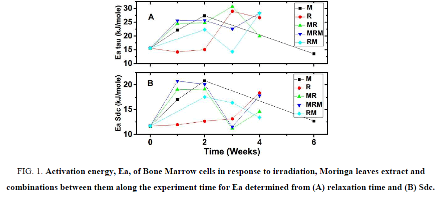
Activation energy, Ea, is determined in this work using two parameters, the dc conductivity (Sdc) and relaxation time of the detected polarisation process. Both parameters were estimated from the fitting of the relaxation process found at a frequency range of (106-4 × 10⁸ Hz) measured by dielectric spectroscopy in our previous work [16].
Moringa leaves extract raised the Ea significantly in the 1st and 2nd week and returned to about the control values, at “0” time, in the 6th week. On the other hand, Irradiation caused Ea to remain about the same in the 1st week and a slight elevation occurred in the 2nd week. In addition, a substantial increase in the 4th week in the activation energy of BM estimated from both relaxation time, FIG. 1A, and dc conductivity, FIG. 1B, of the mentioned relaxation process. The 3rd week had a large elevation in Ea determined by the relaxation time while it showed a slight boost for the Ea estimated from dc conductivity. From the literature it is known that moringa leaves extract is safe for administration up to 5 gm/kg, i.e. showed no toxicity [8,14]. While irradiation with such a high dose, 7.0 ± 0.03 Gy is harmful. Looking at FIG. 1 A and B one can see from the activation energy behaviour along the experimental time that the moringa administration has changed the way by which bone marrow responds radiation damage.
Figure 1: Activation energy, Ea, of Bone Marrow cells in response to irradiation, Moringa leaves extract and combinations between them along the experiment time for Ea determined from (A) relaxation time and (B) Sdc.
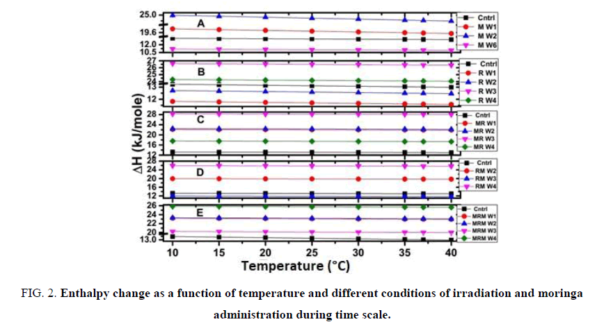
ΔH under the effect of moringa leaves extract, FIG. 2A, is elevated gradually in the 1st and 2nd weeks then descended below the control level in the 6th week.
Figure 2: Enthalpy change as a function of temperature and different conditions of irradiation and moringa administration during time scale.
Radiation caused a decrement of ΔH in the 1st week and started to rise in the 2nd week, still below the control level. ΔH levels high about two folds of the control values in the 3rd week, at last, it decreases in the 4th week towards the control, FIG 2B.
The protected group, MR, in FIG. 2C indicated that moringa effects overwhelmed those of radiation in the 1st week where ΔH rises opposite to the irradiated group. The values of ΔH remained the same during the 2nd week as well confirming the dominance of moringa effect with lesser power. From FIG. 2C it is seen that the 3rd week is nearly the same as the irradiated group and the moringa extract seems to cease its effect, however, the 4th week is closed to the control values than that of the irradiated group which may refer to better healing.
The treated group, RM, FIG. 2D displays a rise in the 2nd week that is less that of M and MR groups indicating Moringa control during this period. The 3rd showed a decrease in ΔH level just below the control, which may refer to the decline of moringa effect. 4th week shows a rise of ΔH similar to that found in the 3rd week of the R group initiating a new period of radiation dominance over the moringa effect in this group which mostly is due to the stopping of moringa administration that lasted for two weeks.
Regarding the protected and protected-treated group, MRM, FIG. 2E it has the MR group’s behavior in the 1st two weeks as ΔH is elevated above the control to the same level. The 3rd week is also elevated above control level having values that are lesser than those of the RM group indicating that moringa effect is continuing up to this stage during the experiment time. Then comes the 4th week which rises just slightly below the level of the 3rd week of the irradiated group shows the evidence of radiation dominance over moringa in the 4th week.
In other words, the moringa leaves extract contradicts the radiation effect with regard to ΔH. In order to reduce the radiation effects, the extract should be given slightly prior to and during the irradiation period and continues after irradiation for as long as the damaging radiation effect is thought to last i.e. in ideal conditions to minimize the radiation injuries as possible, irradiation should be situated at the end of the 1st fourth of moringa leaves extract gavage period to the irradiated subject. Moringa provides protection in the 1st two weeks which are critical and can then reduce mortality.
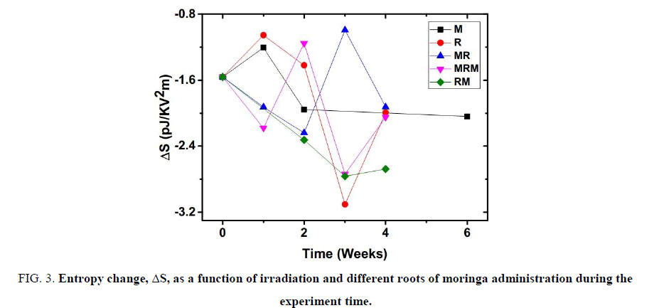
As seen in FIG. 3, Moringa leaves extract as well as irradiation delivers an amount of energy to bone marrow cells. This is shown by the increase of entropy change, ΔS, as its value is moving nearer to zero on the Y-axis. Continual of MOLE feeding led to a decrease of the entropy change that is more than that of irradiation effect getting the bone marrow cells into a more ordered state. The interesting thing noted here, during the two weeks of MOLE administration the effect of MOLE seems to reverse the radiation effect on the entropy change in spite of the resemblance of the MOLE and radiation effects in the 1st week considering each one independently. Only the MRM group deviates in the second week which may be attributed to the submission of the biosystem to MOLE for four weeks along with irradiation after two weeks of MOLE gavage making a huge stress on the biosystem. MOLE administration was stopped after two weeks, the time beyond this period reports the response of different groups and recovery from irradiation in the absence of MOLE. Finally, the change in entropy needs a more detailed study to figure out the bone marrow response using a shorter time scale to map this response and be able to determine the possibility of whether or not to use it as in diagnosis and/or prognosis.
Figure 3: Entropy change, ΔS, as a function of irradiation and different roots of moringa administration during the experiment time.
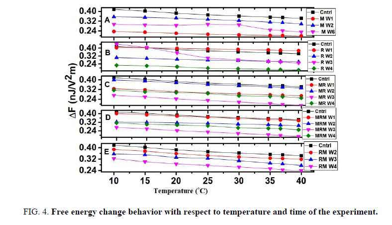
Helmholtz free energy change data had one major difference situated in values of the 1st week. It was markedly decreased for the moringa group, M, FIG. 4A and increased above control values for the irradiated group, R, FIG. 4B. MR group, FIG. 4C, decreased the 1st week, in addition, the 2nd week raised to values just below that of the control. RM group, FIG. 4E, has decreased all measured weeks below the control values.
Figure 4: Free energy change behavior with respect to temperature and time of the experiment.
From the results presents for the change of Helmholtz free energy it is better to use this parameter in the 1st week as there is no much difference for BM cells during the remaining experimental time.
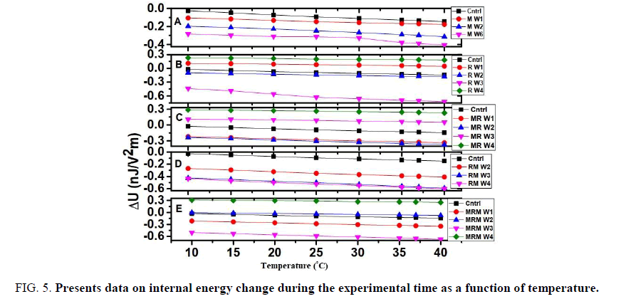
FIG. 5A shows the use of Moringa administration (M group) results in a decrease, along with the Y-axis, of internal energy change up to the 6th week with respect to the control values. Irradiation, R group, caused elevation of the internal energy values in the 1st and 4th weeks while there were decrements in the internal energy values in the 2nd and 3rd weeks, FIG. 5B. FIG. 5C MR group, fed with moringa extract for two weeks prior irradiation, was able to hold the 1st and 2nd weeks below the control level then the 3rd and 4th weeks were raised above the control values. RM group administrated moringa extract for two weeks after irradiation, this group as illustrated in FIG. 5E succeeded to keep the change in internal energy for all the measured weeks below that of control. MRM group, FIG. 5D, MRM one which served moringa extract for two weeks prior and another two weeks after irradiation, could control the 1st and 3rd weeks as for the 2nd week it was raised just above the control values. MRM group failed to control the 4th week which rose above the control level.
Figure 5: Presents data on internal energy change during the experimental time as a function of temperature.
Data refer to that the RM group was the most powerful one as it could maintain the values of the internal energy change during the experimental period below the control level which is the nearest group to the moringa fed un-irradiated group.
Conclusion
During the administration of MOLE, the Ea is increased while irradiation decreased it i.e. made BM cells excited. The same occurs for entropy in spite that MOLE alone has increased entropy change in the 1st week. All other groups cause entropy changes that reverse the direction caused by irradiation except for MRM group in the 2nd week which we think it might be due to the stress caused by long MOLE and irradiation together on the biosystem. Irradiation caused changes in ΔF were reversed by the MOLE administration. Internal energy changes were not as indicative as other parameters. MOLE reverses radiation actions in the 1st two weeks for enthalpy as well which are critical and can then reduce mortality. These results prove that MOLE administration had a positive effect on the biosystem in radiation damage reduction.
Conflicts of Interest
The authors declare no conflicts of interest.
Acknowledgment
The authors would like to thank the STDF project number 2238 for the financial support to buy the KeySight Instrument.
References
- Travlos GS. Normal structure, function, and histology of the bone marrow. Toxicol. Pathol. 2006;34(5):548-65.
- Alter BP. Diagnosis, genetics, and management of inherited bone marrow failure syndromes. ASH Education Program Book. 2007;2007(1):29-39.
- Murakami S, Yoshino H, Ishikawa J, et al. Effects of ionizing radiation on differentiation of murine bone marrow cells into mast cells. J. Radiat. Res. 2015;56(6):865-71.
- Devi PU, Ganasoundari A, Rao BS, et al. In vivo radioprotection by ocimum flavonoids: survival of mice. Radiat Res. 1999;151(1):74-8.
- Samarth RM. Protection against radiation induced hematopoietic damage in bone marrow of Swiss albino mice by Mentha piperita (Linn). J. Radiat. Res. 2007;48(6):523-8.
- Assayed ME, El-Aty AA. Protection of rat chromosomes by melatonin against gamma radiation-induced damage. Mutat Res Genet Toxicol Environ Mutagen. 2009;677(1):14-20.
- An YS, Lee E, Kang MH, et al. Substance P stimulates the recovery of bone marrow after the irradiation. J. Cell. Physiol. 2011;226(5):1204-13.
- Rao AV, Devi PU, Kamath R. In vivo radioprotective effect of Moringa oleifera leaves. Indian J. Experl Biol. 39:858-63.
- Sinha M, Das DK, Bhattacharjee S, et al. Leaf extract of Moringa oleifera prevents ionizing radiation-induced oxidative stress in mice. J Med food. 2011;14(10):1167-72.
- Sinha M, Das DK, Datta S, et al. Amelioration of ionizing radiation induced lipid peroxidation in mouse liver by Moringa oleifera Lam. leaf extract. Indian J. Exp. Biol. 50: 209–15.
- Elwan AM, Salama AA, Said AE, et al. Biophysical and biochemical roles of Moringa oleifera leaves as radioprotector. Prog Biophys Mol Biol. 2018.
- Moyo B, Masika PJ, Hugo A, et al. Nutritional characterization of Moringa (Moringa oleifera Lam.) leaves. Afr. J. Biotechnol. 2011;10(60):12925-33.
- Gopalakrishnan L, Doriya K, Kumar DS. Moringa oleifera: A review on nutritive importance and its medicinal application. Food Sci. Hum. Wellness. 2016;5(2):49-56.
- Okechukwu PU, Okwesili FN, Parker EJ, et al. Phytochemical and acute toxicity studies of Moringa oleifera ethanol leaf extract. Int. J. Life. Sc. Btand Pharm. 2013;2(2):66-71.
- Ezejindu D, Udemezue O, Chinweife K. Hepatoprtective effects of Moringa oleifera extract on liver of wistar rats. Inter. J Res Med Health Sci. 3 (5): 23-27.
- Silva MF, Nishi L, Farooqi A, et al. The many health benefits of Moringa oleifera. J Med Pharm. Innov. 2014;1:9-12.
- Elwan AM, Salama AA, Sayed AM, et al. Response of rats to dose rates of ionizing radiation evaluated by dielectric properties of bone marrow. Prog Biophys Mol Biol. 2018.
- Elnasharty MM, Shousha HA, Elwan AM. Permittivity, entropy, free and internal energies as tools for detecting radiation dose rates’ changes in Hb. Prog Biophys Mol Biol. 2018.
- Elnasharty MM, Ghoneim AM, Elwan AM. Dielectric and thermodynamic study of Hb exploring Moringa oleifera leaves extract potential against radiation damage. Prog Biophys Mol Biol. 2018.
- Płowaś I, Świergiel J, Jadżyn J. Electrical conductivity in dimethyl sulfoxide+ potassium iodide solutions at different concentrations and temperatures. J Chem Eng Data. 2014;59(8):2360-6.
- Haynie DT. The first law of thermodynamics. Biological thermodynamics. Cambridge University Press; 2008: 25-53.
- Jadżyn J, Czechowski G. Prenematic behavior of the electric-field-induced increment of the basic thermodynamic quantities of isotropic mesogenic liquids of different polarity. J Phys Chem. 2007;111(14):3727-9.
- Jadzyn J, Déjardin J, Czechowski G. Singular pretransitional behavior of the electric field-dependent part of the thermodynamic quantities of strongly polar mesogenic liquids in the isotropic phase. Acta Physica Polonica A. 2007;111(6):877.
- Elnasharty MM, Ghoneim AM, Turky GM, et al. Conductivity of cell membrane investigated by a novel dielectrophoretic technique. BioTechnol Ind J. 2013;7(2):60-70.
