Original Article
, Volume: 14( 3)Effect of the Cuttlefish (Sepia officinalis) Pure Ink and the Muscle Extract on Cholestrol Level of Wister Rats Consuming a High Fat Diet: Biotechnological Application
- *Correspondence:
- Benchegra-Rabhia K, Aquaculture and Bioremediation Laboratory (AQUABIOR) – Department of Biotechnology, University of Oran, Ahmed Benbella, Oran, Algeria, Tel: +213 797 08 56 02; E-mail: benchegra.khadidja@yahoo.fr
Received: April 20, 2018;; Accepted: May 04, 2018; Published: May 08, 2018
Citation: Benchegra-Rabhia K, Zouggar AM, Dida-Taleb N, et al. Effect of the Cuttlefish (Sepia officinalis) Pure Ink and the Muscle Extract on Cholestrol Level of Wister Rats Consuming a High Fat Diet: Biotechnological Application. Biotechnol Ind J. 2018;14(3):163.
Abstract
Fisheries co-products are relatively high in some natural biological active components. The presented study attempts to investigate on the effect of natural pure ink and the muscle extract of the cuttlefish (Sepia officinalis) on Cholestrol level of Wistar Rats consuming a high fat diet. The effects of the nutrient intake lipid rich on hypercholesterolemia was determined by a 30-day experiment, in 24 male rats of Wistar strain, aged six weeks and weighing about 200 g. The observed rats are divided into 4 homogeneous groups of six (no: 6); one group receives throughout the experiment a commercial standard diet; the other 03 groups are subjected to a ten-day adaptation phase with a standard commercial diet supplemented with lipids (30% animal fat) (high fat diet: R-HL). After a period of 30 days experiment, a group still consumed the same diet R-HL, the 2nd observed group consumed the HM R-HL supplemented with 250 mg/kg of muscle hydrolysate cuttlefish, and the 3rd EP group consumes the R-HL supplemented with 250 mg/kg pure ink of the cuttlefish. At the 30th date of experiment, the results have shown that the R-HL supplementation of HM and EP has no effect on the contents of total lipids in serum and liver in four groups of rats. However, it modifies their contents in different lipids (total cholesterol, triglycerides, phospholipids). They also cause a 20% increase in the levels of serum proteins in the HM group compared to the HL group. Furthermore, assessment of lipid peroxidation by measuring the TBARS in serum and liver reveal a reduction of 50% in the HM group compared to the HL group. It is observed that some of bioactive components in muscle hydrolysates and pure ink of the cuttlefish (PUFA and bioactive peptides) studied possess considerable metabolic activities (anti-inflammatory and antioxidant). In conclusion, the study claims that the effect of Muscle Hydrolisate Supplementation (MHS) and natural pure ink of the cuttlefish (Sepia officinalis) have a significant effects on lowering Cholestrol level, which will be interesting for an industrial development, for their role in prevention against cardiovascular complications associated with hypercholesterolemia.
Keywords
Cuttlefish; By-product; Cuttlefish muscle hydrlostate; Pure ink; Rat Westar; High fat diet
Introduction
The fishing sector is one of the most important sectors of food production according to the FAO [1]. The cuttlefish, who offers by its exploitation an opportunity of valuation, it produces more than 60% of by-products which turn out relatively rich in certain compound biologically active : essential proteins, amino acids, polyunsaturated fatty acids, enzymes, biopolymers and biomaterials [2]. So, it seems interesting to adopt a strategy of recovery of these substances and to see if it is possible to test them experimentally and to introduce them into the food human or animal as the supplements or the food complements. At present the scientists in search of solutions for one of the most frequent diseases which is Hyperlipidemia or a high level of serum triacylglycerol’s and cholesterol is a risk factor for premature atherosclerosis and coronary heard diseases (CHD). Strong evidences have been put forward by various investigators for the involvement of free radicals production and lipid peroxidation in the onset of atherosclerosis [3].
In Algeria, More than 58% of the deaths are from this disease. The specialists, asserting that more than half death are owed to the cardiovascular diseases, responsible for the death of an Algerian on four that is more than 40000 deaths recorded every year.
A number of epidemiological studies conducted during recent years have clearly demonstrated a link between stress and the development and the course of many diseases [4]. Antioxidants are important aspect of health maintenance based on their modulation of the antioxidative process in the body [5]. Feeding antioxidants attenuate the atherogenic process in animal models, mainly due to their free radical scavenging capabilities [6].
Fish is one of the main components of a healthy diet and many epidemiologic studies and clinical trials have indicated its beneficial effects in incidence of coronary diseases, by decreasing coronary heart disease (CHD) mortality risk.
Many studies from a variety of countries have also reported that seafood consumption protects against lifestyle-related diseases. Numerous epidemiological studies have examined the relationship between dietary marine products and cardiovascular disease (CVD) [7]. In one report, individuals who consumed fatty fish had a 34% reduction in CVD in a three-cohort study [8], and 35 g/day of fish consumption resulted in decreased CVD mortality [9]. A meta-analysis revealed that individuals who consumed fish once a week had a 15% lower risk of CVD mortality compared with individuals who consumed no fish [10]. A previous study suggested that dietary fish protein decreased serum cholesterol through the inhibition of cholesterol.
In vitro studies and the animal model showed antioxidant properties, anti-hypertensives, anti-thrombotic, and bioactive peptides of fishes [11-13]. The aim of the study is to show the effect of supplementations in ink and of muscle hydrolysate on the prevention of high cholesterol level, to the Wistar consuming a high fat diet
Materials and Methods
Preparation of cuttle fish samples
Cuttlefish (Sepia officinalis) were obtained from fish market at Oran City, Algeria. The samples were packed in polyethylene bags, placed in ice and transported to the research laboratory. Upon arrival, it was washed twice with water to eliminate waste. Cuttlefish by-products were separated and only cuttlefish muscle and ink were collected and then stored in sealed plastic bags at -20°C until used for extraction.
Preparation of the lyophilized ink of cuttlefish: The cuttlefish ink lyophilization consists of cold drying process by a lyophilizator. For a long conservation, the dehydration method preserves the nutritional quality of products and the biological properties of molecules. Before the lyophilization the ink was preleved carefully from the ink gland.
Enzymatic hydrolysis of cuttlefish muscle: 100 g of the cuttlefish muscle are solubilizing in 200 ml of distilled water then homogenized during 10 min, the obtained homogenate was heated under agitation during 20 min at 90°C. Then the Protease was added to the mixture (3 U/mg of proteins) and maintained during 6 hours under agitation, at 37°C and pH 7.5 by the addition of a solution of NaOH 4N. After that, the homogenate was then heated at 80°C during 20 min to stop the enzymatic reaction, the mixture was then centrifuged at 5000 g during 20 minutes and the soluble fraction of the cuttlefish proteins hydrolysis was then lyophilized, it was stored at ambient temperature until further use [14].
Animals and diets
Male Wistar rats (Pasteur Institute, Algiers, Algeria) (n=24) weighing 200 ± 15 g were housed in stainless steel cages at temperature of 24°C, with a 12-hours light/dark cycle and relative humidity of 60%. All the rats received a standard diet containing 30% of animal fat, for 10 days. After this adaptation phase, cholesterolemia value was greater and hypercholesterolemic rats were divided into 4 groups (n=6): a group consumed the same diet R-HL, the 2nd observed group consumed the HM R-HL supplemented with 250 mg/kg of muscle hydrolysate cuttlefish, and the 3rd EP group consumes the R-HL supplemented with 250 mg/kg pure ink of the cuttlefish. . Food and tap water were provided ad libitum. Food consumption and weight gain were measured once a week. All animal procedures were in strict accordance with the NIH (No. 85-23, revised 1985) Guide for the Care and Use of Laboratory Animals and all experiments have been examined and approved by our institutional committee for animal care and use (UOB 314/2012).
Blood and tissue samples
After 4 weeks of the experiment and overnight fasting, anaesthetised with chloral (60 mg/kg body weight) and euthanized with an overdose. Blood was collected from abdominal aorta into dried tubes and centrifuged at 4°C, 1000 g for 15 min. Serum was taken and conserved with addition of 0,1% EDTA solution, and separated cells was then washed 3-times by suspending in 0.9% NaCl solution and repeating the centrifugation. The washed cells were lysed in an equal volume of cold water and mixed thoroughly. Liver, muscle were quickly excised in ice-cold saline, blotted on filter paper and weighed. Serum and tissues samples were stored at -70°C until use.
Isolation and characterization of serum LDL-HDL1, HDLM2 and HDL3
Serum LDL-HDL1 was isolated by precipitation using MgCl2 and phosphotungstate (Sigma Chemical Company, France) by the method of Burstein et al. [15]. HDL2 and HDL2 were performed by differential dextran sulphate magnesium chloride precipitation according to Burstein et al. [16]. To estimate the validity of this method, ultracentrifugation was performed according to Havel et al. Total cholesterol (TC) of serum and each fraction was determined by enzymatic colorimetric method (kit Human, GmbH, Wiesbaden, Germany) [17].
Cholesterol determination in serum and hepatic tissue
The total cholesterol is measured by a colorimetric enzymatic method (kit Chronolab Switzerland, Zoug, Switzerland).
Triglyceride determination in serum and hepatic tissue
Triglycerides are determined by a colorimetric enzymatic method (kit Chronolab Switzerland, Zoug, Switzerland). The present TG in the sample gives after enzymatic hydrolysis and oxidation, a quantifiable colored complex by spectrophotometry.
Phospholipids determination in serum and hepatic tissue
Phospholipids are determined by a colorimetric method based on the formation of a colored complex between phospholipids and ammonium ferrothiocyanate.
Lipid peroxidation
Thiobarbituric Acid Reactive Substances (TBARS) concentrations of plasma lipoprotein were measured. One milliliter of lipoprotein fraction was added to 2 mL of thiobarbituric acid (TBA) (final concentration, 0.017 mmol/L), plus butylated hydroxytoluene (concentration, 3.36 µmol/L) and incubated for 15 min at 100°C. After cooling and centrifugation, the absorbance of the supernatant was measured as 535 nm.
Lipid peroxidation in tissue was assessed by the complex formed between malondialdehyde and thiobarbituric acid (TBA). Briefly, liver, aorta tissues (0.5 g) were homogenized with 4.5 mL of KCl (1.15%). The homogenate (100 µL) was mixed with 0.1 mL sodium dodecylsulfate (SDS) (8.1%), 750 µL acetic acid (20%) and 750 µL TBA reagent (0.8%). The reaction mixture was heated at 85°C for 30 min. After heating, the tubes were cooled and 2.5 mL of n-butanol-pyridine (15:1) was added. After mixing and centrifugation at 4000 g for 10 min, the upper phase was taken for measurement at 532 nm [18].
Statistical analysis
The statistical analyses were realized by the software SPSS 17.0 for Windows (SPSSInc., Chicago, III., the USA). All quantitative measurements were expressed as means ± standard deviations (SD) for six rats/group. Data were analyzed using the Tukey test (P 0,05).
The Chi-2 test was used to demonstrate the significant statistical difference (P 0,05) between the groups of animals.
Results
Body weight (BW) and relative organ weights
After four weeks of experiment, a similar body weight was noted in the 4 groups of rats. No significant difference is observed in the evolution the food taking between four groups of animals.
Plasma and tissue lipid concentrations
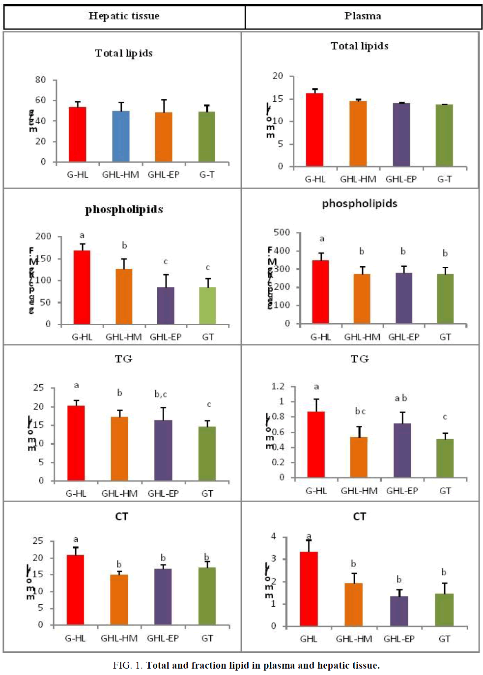
Plasma and hepatic level of total lipids shows no significant difference for the four groups, however at the plasma level compared with the control group the total cholesterol (CT) is significantly decreased in 61% to G-EP and 43% to the G-HM,
The difference between these last two groups and G-T is not significant. The CT is significantly increased by 56% to the G-HL with regard to G-T. At the hepatic level a significant decrease of the contents in CT is noted (28%) for the G-HM and 19% for the G-EP while a not significant difference is noted between these two last ones group and G-T. However a significant increase of 18% in CT is observed at the G-HL with regard to G-T.
Phospholipides at the serum level is decreased in a significant way at the G-HM and the G-EP with regard to (compared with) the G-HL this decrease represents more than 20% to the G-HM and G-EP, while no significant difference is noted between the GT and the G-HM and G-EP. A significant difference is noted between G-T and the G-HL translates by a 22% increase for the G-HL. At the hepatic level a 50% decrease and 26% is noted between the G-HL and the G-EP and the G-HM respectively, and a significant difference between the G-HM and the G-EP, G-T is so noted a significant increase is noted at the G-HL with regard to (compared with) the GT.
The plasma phospholipids level is decreased in a significant way at the G-HM and the G-EP compared with the G-HL this decrease represents more than 20% to the G-HM and G-EP, while no significant difference is noted between the GT and the G-HM and G-EP. A significant difference is noted between G-T and the G-HL (22%) increase for the G-HL. At the hepatic level a 50% decrease and 26% is noted between the G-HL and the G-EP and the G-HM respectively, and a significant difference between the G-HM and the G-EP, G-T is so noted also a significant increase is noted at the G-HL compared with the GT.
The plasma triglyceride (TG) a significant decrease of 38% is noted between the G-HL and G-HM, and a not significant decrease of 17% is noted at the G-EP. The difference between both Handled G-HL presents no significant difference while the TG to the G-EP is significantly raised compared to the GT however the rate of TG is significantly increased 41% to the G-HL compared with the GT. At the hepatic level a significant decrease of the TG of the order of 14% and 18% for the G-HM and G-EP respectively with regard to the G-HL and the increase for this last group compared to G-T and of 27%, the rate of TG is significantly higher at the G-HM compared to the GT, the difference between the GT and the G-EP is not significant (FIG. 1).
Triglycerides and total cholesterol repartition in lipid fractions
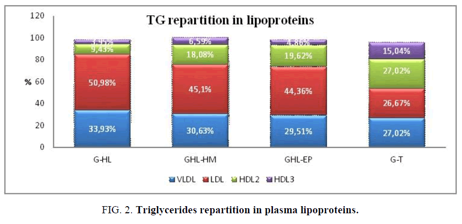
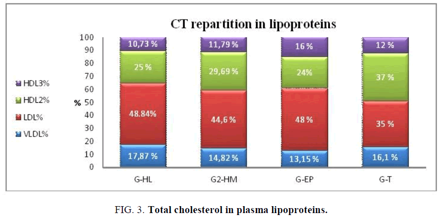
The results show that the plasma TG is distributed in a similar way between the groups HL, HM EP, and more than 40% of the plasma TG is carried by the LDL, however the difference not significant between the proportions of various fractions of lipoproteins is observed at the group T. The plasma CT is mainly carried by the VLDL and the LDL to four groups and no significant difference is noted between 4 groups in the repartition of the proportions of the CT at the level of lipoproteins (FIG. 2).
Lipid peroxydation levels
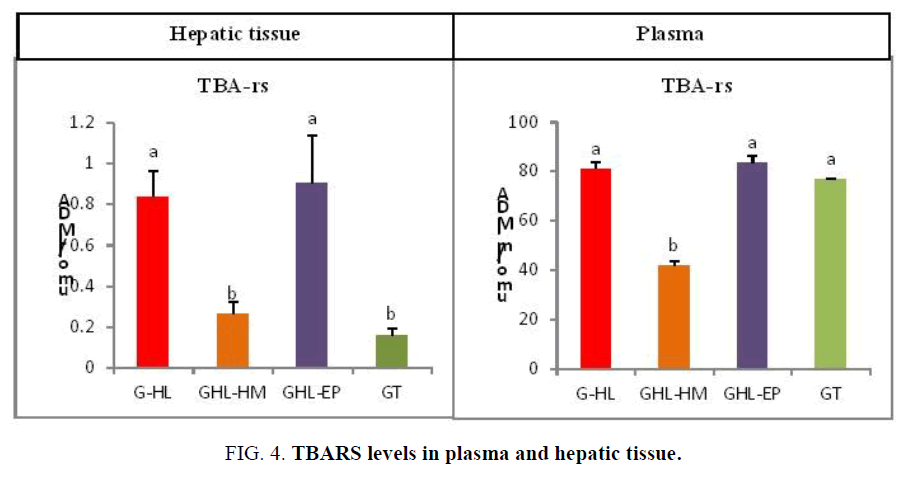
At the plasma level the TBARS contents were significantly decreased at the group HM with compared with the other groups, however in the hepatic tissue the TBARS were significantly higher in the group HL compared with the GT (FIG. 3 and FIG. 4).
In the group HM the TBARS were significantly decreased and do not represent a significant difference with the GT. However no significant decrease is noted at the group EP compared with the group HL.
Discussion
The objective of this experimental study is to highlight the power of the cuttlefish muscle hydrolysat and pure ink to reduce cholesterol of the rat submitted to a high fat diet. After J30 of experiment the result indicate that HM and EP had effect neither on the taking food nor on the weight of animals. Also the relative weight of the liver is not influenced. These results are in agreement with those of Hosomi et al. [19], which observed no difference in the animal’s weight, the taking food or the relative weight of the liver and the adipose tissue by comparing between two subdued groups, one containing 10% of Alaska hake peptides and 10% of casein and the other 20% of casein.
The rate of plasma cholesterol is a major risk factor of the cardiovascular diseases. The rise of the rate of cholesterol is the only one to be able to lead the development of atheroma plate, even in the absence of other risk factors. The modification of the rates of plasma lipids can influence the composition, the content and the distribution of the subclasses of plasma lipoproteins. Our results do not show an accumulation of total lipids at the hepatic tissue and plasma.
However the contents in hepatic total cholesterol are decreased at the group HM and particularly at the group EP. These decreases in the rate of the CT could be attributed to the bioactive peptides.
Similar studies showed that the fish hydrolysat act as regulators of the cholesterol metabolism, in the study of Hosomi et al [19], a decrease of the contents in hepatic and plasma CT is observed at rats subjected during four weeks has a diet of Alaska hake hydrolysat, more recently the same authors [19] noted that a contribution it is thihydrolysat decreases the serum and hepatic contents in CT to the rat Wistar consuming a hypercholesterolemic diet. Also, Wergedahl et al. [20] report that a diet of salmon hydrolysat after twelve days to the rat Wistar a decrease of the cholesterol level by comparison to the control group.
After four weeks a high level of triglycerides is noted at the group HL compared with the group, due to the supplementations of fat (30%) in the diet. After 30 days of supplemented in HM and EP, the values of the plasma TG are reduced and get closer to those of the GT. These results are in agreements with those of Ben khaled et al. [14] and Liaset et al. [21].
In this study, the concentrations in plasma phospholipides the liver are decreased compared to the group HM and the group EP. This decrease of the PL results from a reduction of the concentrations in PL at the level of the VLDL and from HDL3 probably owed to their low synthesis by the liver.
Our results indicate that at plasma, the decrease of the contents in total cholesterol in the muscle hydrolisate and the pure ink was mainly due to the low contents in C-VLDL and C-LDL. These results join those of Ben Khaled et al. [14]] and Hosomi et al. [19] who noted a decrease of the cholesterol in the fraction LDL. In this study, the decrease of the C-VLDL and the C-LDL in answer to the treatment with the EP and the HM mainly reflects partially the low concentrations of the hepatic cholesterol.
The liver is involved in the metabolism of the VLDL. The decrease or the increase of the contents in hepatic triglycerides generates impoverished VLDL or enrich in TG. The content in similar TG-VLDL lets suggest, an identical hepatic synthesis of the TG or a catabolism of fatty acids.
Our results concerning the TBARS in plasma are in agreement with those of Durak et al. [22] who note that an excessive contribution in cholesterol does not seem to affect the antioxidative activity mainly antioxidative enzymes represented by superoxide dismutase, the catalase and the glutathion peroxydase with the exception of the activity paraoxonase which is decreased.
While at the hepatic level our results join those of Anilla and Vijiyalakshmi [23], who notes that a diet rich in cholesterol affects the antioxidative system to the animal further to an increase of the peroxydation of lipids and formation of the free radicals.
The studies of Erdmann et al. [11] Shahidi and Zhong suggest that the protein hydrolysats could act as chelators of metals, or the inhibitors of the lipid peroxyation.
Ben khaled et al. [14]] noticed in their work that adipeptide (Met-Tyr) stemming from the hydrolyase of proteins of Sardine stimulates the expression of two antioxidative proteins in cells what has for consequence a protection against the oxidation responsible for the attack of the polyunsaturated fatty acids.
The antioxidative activity of the bioactive peptides would also be awarded according to Erdmann et al. [11] Shahidi and Zhong in hydrophobic amino acids which could act as donors of proton of their function hydroxylates as the case of most part of antioxidants.
If many works studied the effect of proteins of fish or their hydrolysat on the lipid peroxydation in the rat consuming a hyperlipid diet no study is concerned the pure ink of the cuttlefish.
Conclusion
In view of its results, the incorporation of the cuttle fish muscle hydrolysat and the pure Ink in a dietary program could be strategically effective to prevent, even improve the dyslipidemia and limit the radicalaire attack, what will allow to prevent the cardiovascular complications associated with the hypercholesterol level.
So, the hydrolysat of the muscle and the ink of the cuttlefish should be valued seen its nutritional and therapeutic remarkable interest. Further clinical studies are needed to elucidate this hypothesis.
References
- FAO. The State of World Fisheries and Aquaculture. 2014.
- Ferraro V, Carvalho AP, Piccirillo C, et al. Extraction of high added value biological compounds from sardine, sardine-type fish and mackerel canning residues-a review. Materials Sci Eng C. 33(6):3111-20.
- Bansal MP, Jaswal S. Hypercholesterolemia induced oxidative stress is reduced in rats with diets enriched with supplement from Dunaliella salina algae. Am J Biomed Sci. 2009;1(3):196-204.
- Gümüslü S, Bilmen Sarikçiog?lu S, Sahin E, et al. Influences of different stress models on the antioxidant status and lipid peroxidation in rat erythrocytes. Free Radic Res. 2002;36(12):1277-82.
- Lee MK, Bok SH, Jeong TS, et al. Supplementation of naringenin and its synthetic derivative alters antioxidant enzyme activities of erythrocyte and liver in high cholesterol-fed rats. Bioorg Med Chem. 2002;10(7):2239-44.
- Paul A, Calleja L, Joven J, et al. Supplementation with vitamin E and/or zinc does not attenuate atherosclerosis in apolipoprotein E-deficient mice fed a high-fat, high-cholesterol diet. Int J Vitamin Nutr Res. 2001;71(1):45-52.
- Guallar E, Sanz-Gallardo MI, Veer PV, et al. Mercury, fish oils, and the risk of myocardial infarction. New Engl J Med. 2002;347(22):1747-54.
- Oomen CM, Feskens EJ, Räsänen L, et al. Fish consumption and coronary heart disease mortality in Finland, Italy, and The Netherlands. Am J Epidemiol. 2000;151(10):999-1006.
- Daviglus ML, Stamler J, Orencia AJ, et al. Fish consumption and the 30-year risk of fatal myocardial infarction. New England J Med. 1997;336(15):1046-53.
- He K, Song Y, Daviglus ML, et al. Accumulated evidence on fish consumption and coronary heart disease mortality: a meta-analysis of cohort studies. Circulation. 2004;109(22):2705-11.
- Erdmann K, Cheung BW, Schröder H. The possible roles of food-derived bioactive peptides in reducing the risk of cardiovascular disease. J Nutr Biochem. 2008;19(10):643-54.
- Möller NP, Scholz-Ahrens KE, Roos N, et al. Bioactive peptides and proteins from foods: Indication for health effects. Eur J Nutr. 2008;47(4):171-82.
- Shahidi F, Zhong Y. Bioactive peptides. J AOAC Int. 2008;91:914-31.
- Khaled HB, Ghlissi Z, Chtourou Y, et al. Effect of protein hydrolysates from sardinelle (Sardinella aurita) on the oxidative status and blood lipid profile of cholesterol-fed rats. Food Res Int. 2012;45(1):60-8.
- Burstein M, Scholnick HR, Morfin R. Rapid method for the isolation of lipoproteins from human serum by precipitation with polyanions. J Lipid Res. 1970;11(6):583-95.
- Burstein M, Fine A, Atger VR, et al. Rapid method for the isolation of two purified subfractions of high density lipoproteins by differential dextran sulfate-magnesium chloride precipitation. Biochimie. 1989;71(6):741-6.
- Gümüslü S, Bilmen Sarikçiog?lu S, Sahin E, et al. Influences of different stress models on the antioxidant status and lipid peroxidation in rat erythrocytes. Free Radic Res. 2002;36(12):1277-82.
- Ohkawa H, Ohishi N, Yagi K. Assay for lipid peroxides in animal tissues by thiobarbituric acid reaction. Anal Biochem. 1979;95(2):351-8.
- Hosomi R, Fukunaga K, Arai H, et al. Effect of simultaneous intake of fish protein and fish oil on cholesterol metabolism in rats fed high-cholesterol diets. Cellulose. 2011;50(50):50.
- Wergedahl H, Liaset B, Gudbrandsen OA, et al. Fish protein hydrolysate reduces plasma total cholesterol, increases the proportion of HDL cholesterol, and lowers acyl-CoA: cholesterol acyltransferase activity in liver of Zucker rats. J Nutr. 2004;134(6):1320-7.
- Liaset B, Madsen L, Hao Q, et al. Fish protein hydrolysate elevates plasma bile acids and reduces visceral adipose tissue mass in rats. Biochim Biophysic Acta. 2009;1791(4):254-62.
- Durak I, Özbek H, Devrim E, et al. Effects of cholesterol supplementation on antioxidant enzyme activities in rat hepatic tissues: Possible implications of hepatic paraoxonase in atherogenesis. Nutr Metab Cardiovasc Dis. 2004;14(4):211-4.
- Anila L, Vijayalakshmi NR. Antioxidant action of flavonoids from Mangifera indica and Emblica officinalis in hypercholesterolemic rats. Food Chem. 2003;83(4):569-74.
